↩ Accueil

Vue normale

Microbubbles power soft, programmable artificial muscles

10 novembre 2025 à 10:30
Ultrasound-powered soft surgical robot
Ultrasound-powered stingraybot A bioinspired soft surgical robot with artificial muscles made from microbubble arrays swims forward under swept-frequency ultrasound excitation. Right panels: motion of the microbubble-array fins during swimming. Lower inset: schematic of the patterned microbubble arrays. Scale bar: 1 cm. (Courtesy: CC BY 4.0/Nature 10.1038/s41586-025-09650-3)

Artificial muscles that offer flexible functionality could prove invaluable for a range of applications, from soft robotics and wearables to biomedical instrumentation and minimally invasive surgery. Current designs, however, are limited by complex actuation mechanisms and challenges in miniaturization. Aiming to overcome these obstacles, a research team headed up at the Acoustic Robotics Systems Lab (ETH Zürich) in Switzerland is using microbubbles to create soft, programmable artificial muscles that can be wirelessly controlled via targeted ultrasound activation.

Gas-filled microbubbles can concentrate acoustic energy, providing a means to initiate movement with rapid response times and high spatial accuracy. In this study, reported in Nature, team leader Daniel Ahmed and colleagues built a synthetic muscle from a thin flexible membrane containing arrays of more than 10,000 microbubbles. When acoustically activated, the microbubbles generate thrust and cause the membrane to deform. And as different sized microbubbles resonate at different ultrasound frequencies, the arrays can be designed to provide programmable motion.

“Ultrasound is safe, non-invasive, can penetrate deep into the body and can generate large forces. However, without microbubbles, a much higher force is needed to deform the muscle, and selective activation is difficult,” Ahmed explains. “To overcome this limitation, we use microbubbles, which amplify force generation at specific sites and act as resonant systems. As a result, we can activate the artificial muscle at safe ultrasound power levels and generate complex motion.”

The team created the artificial muscles from a thin silicone membrane patterned with an array of cylindrical microcavities with the dimensions of the desired microbubbles. Submerging this membrane in a water-filled acoustic chamber trapped tens of thousands of gas bubbles within the cavities (one per cavity). The final device contains around 3000 microbubbles per mm2 and weighs just 0.047 mg/mm2.

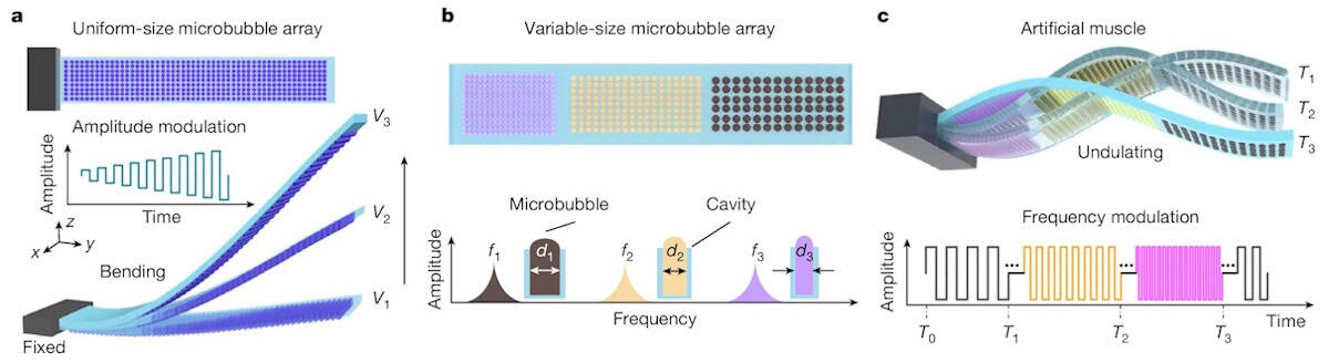
To demonstrate acoustic activation, the researchers fabricated an artificial muscle containing uniform-sized microbubbles on one surface. They fixed one end of the muscle and exposed it to resonant frequency ultrasound, simultaneously exciting the entire microbubble array. The resulting oscillations generated acoustic streaming and radiation forces, causing the muscle to flex upward, with an amplitude dependent upon the ultrasound excitation voltage.

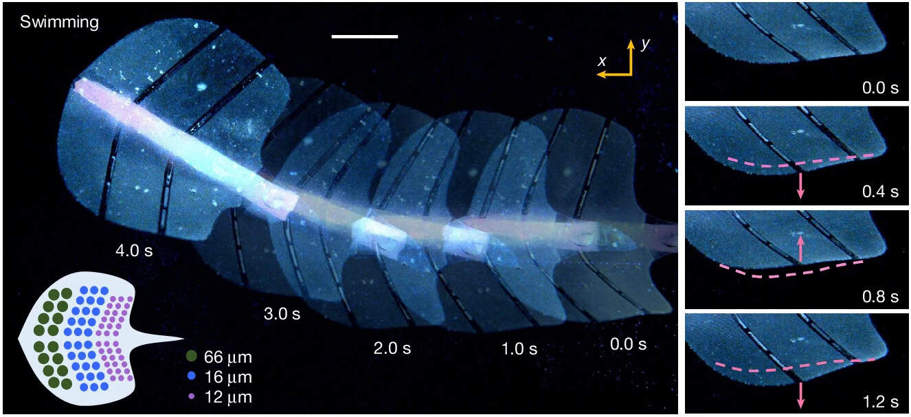
Next, the team designed an 80 µm-thick, 3 x 0.5 cm artificial muscle containing arrays of three different sized microbubbles. Stimulation at 96.5, 82.3 and 33.2 kHz induced deformations in regions containing bubbles with diameters of 12, 16 and 66 µm, respectively. Exposure to swept-frequency ultrasound covering the three resonant frequencies sequentially activated the different arrays, resulting in an undulatory motion.

Microbubble-array artificial muscles
Microbubble muscles (a) Artificial muscle with thousands of microbubbles on its lower surface bends upwards when excited by ultrasound. (b) Artificial muscle containing arrays of microbubbles with three different diameters, each corresponding to a distinct natural frequency, exhibits undulatory motion (c) under swept-frequency ultrasound excitation. (Courtesy: CC BY 4.0/Nature 10.1038/s41586-025-09650-3)

A multitude of functions

Ahmed and colleagues showcased a range of applications for the artificial muscle by integrating microbubble arrays into functional devices, such as a miniature soft gripper for trapping and manipulating fragile live animals. The gripper comprises six to ten microbubble array-based “tentacles” that, when subjected to ultrasound, gently gripped a zebrafish larva with sub-100 ms response time. When the ultrasound was switched off, the tentacles opened and the larva swam away with no adverse effects.

The artificial muscle can function as a conformable robotic skin that sticks and imparts motion to a stationary object, which the team demonstrated by attaching it to the surface of an excised pig heart. It can also be employed for targeted drug delivery – shown by the use of a microbubble-array robotic patch for ultrasound-enhanced delivery of dye into an agar block.

The researchers also built an ultrasound-powered “stingraybot”, a soft surgical robot with artificial muscles (arrays of differently sized microbubbles) on either side to mimic the pectoral fins of a stingray. Exposure to swept-frequency ultrasound induced an undulatory motion that wirelessly propelled the 4 cm-long robot forward at a speed of about 0.8 body lengths per second.

To demonstrate future practical biomedical applications, such as supporting minimally invasive surgery or site-specific drug release within the gastrointestinal tract, the researchers encapsulated a rolled up stingraybot within a 27 x 12 mm edible capsule. Once released into the stomach, the robot could be propelled on demand under ultrasound actuation. They also pre-folded a linear artificial muscle into a wheel shape and showed that swept ultrasound frequencies could propel it along the complex mucosal surfaces of the stomach and intestine.

“Through the strategic use of microbubble configurations and voltage and frequency as ultrasound excitation parameters, we engineered a diverse range of preprogrammed movements and demonstrated their applicability across various robotic platforms,” the researchers write. “Looking ahead, these artificial muscles hold transformative potential across cutting-edge fields such as soft robotics, haptic medical devices and minimally invasive surgery.”

Ahmed says that the team is currently developing soft patches that can conform to biological surfaces for drug delivery inside the bladder. “We are also designing soft, flexible robots that can wrap around a tumour and release drugs directly at the target site,” he tells Physics World. “Basically, we’re creating mobile conformable drug-delivery patches.”

The post Microbubbles power soft, programmable artificial muscles appeared first on Physics World.

Ultrasound probe maps real-time blood flow across entire organs

6 novembre 2025 à 10:35

Microcirculation – the flow of blood through the smallest vessels – is responsible for distributing oxygen and nutrients to tissues and organs throughout the body. Mapping this flow at the whole-organ scale could enhance our understanding of the circulatory system and improve diagnosis of vascular disorders. With this aim, researchers at the Institute Physics for Medicine Paris (Inserm, ESPCI-PSL, CNRS) have combined 3D ultrasound localization microscopy (ULM) with a multi-lens array method to image blood flow dynamics in entire organs with micrometric resolution, reporting their findings in Nature Communications.

“Beyond understanding how an organ functions across different spatial scales, imaging the vasculature of an entire organ reveals the spatial relationships between macro- and micro-vascular networks, providing a comprehensive assessment of its structural and functional organization,” explains senior author Clement Papadacci.

The 3D ULM technique works by localizing intravenously injected microbubbles. Offering a spatial resolution roughly ten times finer than conventional ultrasound, 3D ULM can map and quantify micro-scale vascular structures. But while the method has proved valuable for mapping whole organs in small animals, visualizing entire organs in large animals or humans is hindered by the limitations of existing technology.

To enable wide field-of-view coverage while maintaining high-resolution imaging, the team – led by PhD student Nabil Haidour under Papadacci’s supervision – developed a multi-lens array probe. The probe comprises an array of 252 large (4.5 mm²) ultrasound transducer elements. The use of large elements increases the probe’s sensitive area to a total footprint of 104 x 82 mm, while maintaining a relatively low element count.

Each transducer element is equipped with an individual acoustic diverging lens. “Large elements alone are too directive to create an image, as they cannot generate sufficient overlap or interference between beams,” Papadacci explains. “The acoustic lenses reduce this directivity, allowing the elements to focus and coherently combine signals in reception, thus enabling volumetric image formation.”

Whole-organ imaging

After validating their method via numerical simulations and phantom experiments, the team used a multi-lens array probe driven by a clinical ultrasound system to perform 3D dynamic ULM of an entire explanted porcine heart – considered an ideal cardiac model as its vascular anatomies and dimensions are comparable to those of humans.

The heart was perfused with microbubble solution, enabling the probe to visualize the whole coronary microcirculation network over a large volume of 120 x 100 x 82 mm, with a spatial resolution of around 125 µm. The technique enabled visualization of both large vessels and the finest microcirculation in real time. The team also used a skeletonization algorithm to measure vessel radii at each voxel, which ranged from approximately 75 to 600 µm.

As well as structural imaging, the probe can also assess flow dynamics across all vascular scales, with a high temporal resolution of 312 frames/s. By tracking the microbubbles, the researchers estimated absolute flow velocities ranging from 10 mm/s in small vessels to over 300 mm/s in the largest. They could also differentiate arteries and veins based on the flow direction in the coronary network.

In vivo demonstrations

Next, the researchers used the multi-lens array probe to image the entire kidney and liver of an anaesthetized pig at the Veterinary school of Maison Alfort, with the probe positioned in front of the kidney or liver, respectively, and held using an articulated arm. They employed electrocardiography to synchronize the ultrasound acquisitions with periods of minimal respiratory motion and injected microbubble solution intravenously into the animal’s ear.

In vivo imaging of a porcine kidney
In vivo imaging Left: 3D microbubble density map of the porcine kidney. Centre: 3D flow map of microbubble velocity distribution. Right: 3D flow map showing arterial (red) and venous (blue) flow. (Courtesy: CC BY 4.0/Nat. Commun. 10.1038/s41467-025-64911-z)

The probe mapped the vascular network of the kidney over a 60 x 80 x 40 mm volume with a spatial resolution of 147 µm. The maximum 3D absolute flow velocity was approximately 280 mm/s in the large vessels and the vessel radii ranged from 70 to 400 µm. The team also used directional flow measurements to identify the arterial and venous flow systems.

Liver imaging is more challenging due to respiratory, cardiac and stomach motions. Nevertheless, 3D dynamic ULM enabled high-depth visualization of a large volume of liver vasculature (65 x 100 x 82 mm) with a spatial resolution of 200 µm. Here, the researchers used dynamic velocity measurement to identify the liver’s three blood networks (arterial, venous and portal veins).

“The combination of whole-organ volumetric imaging with high-resolution vascular quantification effectively addresses key limitations of existing modalities, such as ultrasound Doppler imaging, CT angiography and 4D flow MRI,” they write.

Clinical applications of 3D dynamic ULM still need to be demonstrated, but Papadacci suggests that the technique has strong potential for evaluating kidney transplants, coronary microcirculation disorders, stroke, aneurysms and neoangiogenesis in cancer. “It could also become a powerful tool for monitoring treatment response and vascular remodelling over time,” he adds.

Papadacci and colleagues anticipate that translation to human applications will be possible in the near future and plan to begin a clinical trial early in 2026.

The post Ultrasound probe maps real-time blood flow across entire organs appeared first on Physics World.

Tumour-specific radiofrequency fields suppress brain cancer growth

28 octobre 2025 à 14:00

A research team headed up at Wayne State University School of Medicine in the US has developed a novel treatment for glioblastoma, based on exposure to low levels of radiofrequency electromagnetic fields (RF EMF). The researchers demonstrated that the new therapy slows the growth of glioblastoma cells in vitro and, for the first time, showed its feasibility and clinical impact in patients with brain tumours.

The study, led by Hugo Jimenez and reported in Oncotarget, uses a device developed by TheraBionic that delivers amplitude-modulated 27.12 MHz RF EMF throughout the entire body, via a spoon-shaped antenna placed on the tongue. Using tumour-specific modulation frequencies, the device has already received US FDA approval for treating patients with advanced hepatocellular carcinoma (HCC, a liver cancer), while its safety and effectiveness are currently being assessed in clinical trials in patients with pancreatic, colorectal and breast cancer.

In this latest work, the team investigated its use in glioblastoma, an aggressive and difficult to treat brain tumour.

To identify the particular frequencies needed to treat glioblastoma, the team used a non-invasive biofeedback method developed previously to study patients with various types of cancer. The process involves measuring variations in skin electrical resistance, pulse amplitude and blood pressure while individuals are exposed to low levels of amplitude-modulated frequencies. The approach can identify the frequencies, usually between 1 Hz and 100 kHz, specific to a single tumour type.

Jimenez and colleagues first examined the impact of glioblastoma-specific amplitude-modulated RF EMF (GBMF) on glioblastoma cells, exposing various cell lines to GBMF for 3 h per day at the exposure level used for patient treatments. After one week, GBMF decreased the proliferation of three glioblastoma cell lines (U251, BTCOE-4765 and BTCOE-4795) by 34.19%, 15.03% and 14.52%, respectively.

The team note that the level of this inhibitive effect (15–34%) is similar to that observed in HCC cell lines (19–47%) and breast cancer cell lines (10–20%) treated with tumour-specific frequencies. A fourth glioblastoma cell line (BTCOE-4536) was not inhibited by GBMF, for reasons currently unknown.

Next, the researchers examined the effect of GBMF on cancer stem cells, which are responsible for treatment resistance and cancer recurrence. The treatment decreased the tumour sphere-forming ability of U251 and BTCOE-4795 cells by 36.16% and 30.16%, respectively – also a comparable range to that seen in HCC and breast cancer cells.

Notably, these effects were only induced by frequencies associated with glioblastoma. Exposing glioblastoma cells to HCC-specific modulation frequencies had no measurable impact and was indistinguishable from sham exposure.

Looking into the underlying treatment mechanisms, the researchers hypothesized that – as seen in breast cancer and HCC – glioblastoma cell proliferation is mediated by T-type voltage-gated calcium channels (VGCC). In the presence of a VGCC blocker, GBMF did not inhibit cell proliferation, confirming that GBMF inhibition of cell proliferation depends on T-type VGCCs, in particular, a calcium channel known as CACNA1H.

The team also found that GBMF blocks the growth of glioblastoma cells by modulating the “Mitotic Roles of Polo-Like Kinase” signalling pathway, leading to disruption of the cells’ mitotic spindles, critical structures in cell replication.

A clinical first

Finally, the researchers used the TheraBionic device to treat two patients: a 38-year-old patient with recurrent glioblastoma and a 47-year-old patient with the rare brain tumour oligodendroglioma. The first patient showed signs of clinical and radiological benefit following treatment; the second exhibited stable disease and tolerated the treatment well.

“This is the first report showing feasibility and clinical activity in patients with brain tumour,” the authors write. “Similarly to what has been observed in patients with breast cancer and hepatocellular carcinoma, this report shows feasibility of this treatment approach in patients with malignant glioma and provides evidence of anticancer activity in one of them.”

The researchers add that a previous dosimetric analysis of this technique measured a whole-body specific absorption rate (SAR, the rate of energy absorbed by the body when exposed to RF EMF) of 1.35 mW/kg and a peak spatial SAR (over 1 g of tissue) of 146–352 mW/kg. These values are well within the safety limits set by the ICNIRP (whole-body SAR of 80 mW/kg; peak spatial SAR of 2000 mW/kg). Organ-specific values for grey matter, white matter and the midbrain also had mean SAR ranges well within the safety limits.

The team concludes that the results justify future preclinical and clinical studies of the TheraBionic device in this patient population. “We are currently in the process of designing clinical studies in patients with brain tumors,” Jimenez tells Physics World.

The post Tumour-specific radiofrequency fields suppress brain cancer growth appeared first on Physics World.

Resonant laser ablation selectively destroys pancreatic tumours

23 octobre 2025 à 10:00

Pancreatic ductal adenocarcinoma (PDAC), the most common type of pancreatic cancer, is an aggressive tumour with a poor prognosis. Surgery remains the only potential cure, but is feasible in just 10–15% of cases. A team headed up at Sichuan University in China has now developed a selective laser ablation technique designed to target PDAC while leaving healthy pancreatic tissue intact.

Thermal ablation techniques, such as radiofrequency, microwave or laser ablation, could provide a treatment option for patients with locally advanced PDAC, but existing methods risk damaging surrounding blood vessels and healthy pancreatic tissues. The new approach, described in Optica, uses the molecular fingerprint of pancreatic tumours to enable selective ablation.

The technique exploits the fact that PDAC tissue contains a large amount of collagen compared with healthy pancreatic tissue. Amide-I collagen fibres exhibit a strong absorption peak at 6.1 µm, thus the researchers surmised that tuning the treatment laser to this resonant wavelength could enable efficient tumour ablation with minimal collateral thermal damage. As such, they designed a femtosecond pulsed laser that can deliver 6.1 µm pulses with a power of more than 1 W.

FTIR spectra of PDAC and the laser
Resonant wavelength Fourier-transform infrared spectra of PDAC (blue) and the laser (red). (Courtesy: Houkun Liang, Sichuan University)

“We developed a mid-infrared femtosecond laser system for the selective tissue ablation experiment,” says team leader Houkun Liang. “The system is tunable in the wavelength range of 5 to 11 µm, aligning with various molecular fingerprint absorption peaks such as amide proteins, cholesteryl ester, hydroxyapatite and so on.”

Liang and colleagues first examined the ablation efficiency of three different laser wavelengths on two types of pancreatic cancer cells. Compared with non-resonant wavelengths of 1 and 3 µm, the collagen-resonant 6.1 µm laser was far more effective in killing pancreatic cancer cells, reducing cell viability to ranges of 0.27–0.32 and 0.37–0.38, at 0 and 24 h, respectively.

The team observed similar results in experiments on ectopic PDAC tumours cultured on the backs of mice. Irradiation at 6.1 µm led to five to 10 times deeper tumour ablation than seen for the non-resonant wavelengths (despite using a laser power of 5 W for 1 µm ablation and just 500 mW for 6.1 and 3 µm), indicating that 6.1 µm is the optimal wavelength for PDAC ablation surgery.

To validate the feasibility and safety of 6.1 µm laser irradiation, the team used the technique to treat PDAC tumours on live mice. Nine days after ablation, the tumour growth rate in treated mice was significantly suppressed, with an average tumour volume of 35.3 mm3. In contrast, tumour volume in a control group of untreated mice reached an average of 292.7 mm3, roughly eight times the size of the ablated tumours. No adverse symptoms were observed following the treatment.

Clinical potential

The researchers also used 6.1 µm laser irradiation to ablate pancreatic tissue samples (including normal tissue and PDAC) from 13 patients undergoing surgical resection. They used a laser power of 1 W and four scanning speeds (0.5, 1, 2 and 3 mm/s) with 10 ablation passes, examining 20 to 40 samples for each parameter.

At the slower scanning speeds, excessive energy accumulation resulted in comparable ablation depths. At speeds of 2 or 3 mm/s, however, the average ablation depths in PDAC samples were 2.30 and 2.57 times greater than in normal pancreatic tissue, respectively, demonstrating the sought-after selective ablation. At 3 mm/s, for example, the ablation depth in tumour was 1659.09±405.97 µm, compared with 702.5±298.32 µm in normal pancreas.

The findings show that by carefully controlling the laser power, scanning speed and number of passes, near-complete ablation of PDACs can be achieved, with minimal damage to surrounding healthy tissues.

To further investigate the clinical potential of this technique, the researchers developed an anti-resonant hollow-core fibre (AR-HCF) that can deliver high-power 6.1 µm laser pulses deep inside the human body. The fibre has a core diameter of approximately 113 µm and low bending losses at radii under 10 cm. The researchers used the AR-HCF to perform 6.1 µm laser ablation of PDAC and normal pancreas samples. The ablation depth in PDAC was greater than in normal pancreas, confirming the selective ablation properties.

“We are working together with a company to make a medical-grade fibre system to deliver the mid-infrared femtosecond laser. It consists of AR-HCF to transmit mid-infrared femtosecond pulses, a puncture needle and a fibre lens to focus the light and prevent liquid tissue getting into the fibre,” explains Liang. “We are also making efforts to integrate an imaging unit into the fibre delivery system, which will enable real-time monitoring and precise surgical guidance.”

Next, the researchers aim to further optimize the laser parameters and delivery systems to improve ablation efficiency and stability. They also plan to explore the applicability of selective laser ablation to other tumour types with distinct molecular signatures, and to conduct larger-scale animal studies to verify long-term safety and therapeutic outcomes.

“Before this technology can be used for clinical applications, highly comprehensive biological safety assessments are necessary,” Liang emphasizes. “Designing well-structured clinical trials to assess efficacy and risks, as well as navigating regulatory and ethical approvals, will be critical steps toward translation. There is a long way to go.”

The post Resonant laser ablation selectively destroys pancreatic tumours appeared first on Physics World.

❌