↩ Accueil

Vue lecture

Metamaterial antennas enhance MR images of the eye and brain

In vivo MR imaging
In vivo imaging T2-weighted MRI of three healthy volunteers (left to right columns) using the bend-MTMA and bend-loop antennas reveals increased intraocular signal for the metamaterial-based bend-MTMA configuration. (Courtesy: CC BY 4.0/Advanced Materials 10.1002/adma.202517760)

MRI is one of the most important imaging tools employed in medical diagnostics. But for deep-lying tissues or complex anatomic features, MRI can struggle to create clear images in a reasonable scan time. A research team led by Thoralf Niendorf at the Max Delbrück Center in Germany is using metamaterials to create a compact radiofrequency (RF) antenna that enhances image quality and enables faster MRI scanning.

Imaging the subtle structures of the eye and orbit (the surrounding eye socket) is a particular challenge for MRI, due to the high spatial resolution and small fields-of-view required, which standard MRI systems struggle to achieve. These limitations are generally due to the antennas (or RF coils) that transmit and receive the RF signals. Increasing the sensitivity of these antennas will increase signal strength and improve the resolution of the resulting MR images.

To achieve this, Niendorf and colleagues turned to electromagnetic metamaterials – artificially manufactured, regularly arranged structures made of periodic subwavelength unit cells (UCs) that interact with electromagnetic waves in ways that natural materials do not. They designed the metamaterial UCs based on a double-square split-ring resonator design, tailored for operation at a high magnetic field strength of 7.0 T.

Metamaterials improve transmit–receive performance

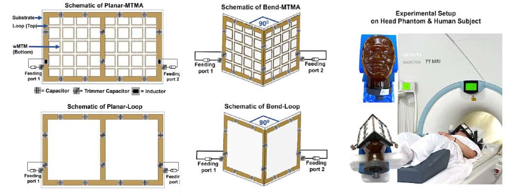
In their latest study, led by doctoral student Nandita Saha and reported in Advanced Materials, the researchers created a metamaterial-integrated RF antenna (MTMA) by fabricating the UCs into a 5 x 8 array. They built two configurations: a planar antenna (planar-MTMA); and a version with a 90° bend in the centre (bend-MTMA) to conform to the human face. For comparison, they also built conventional counterparts without the metamaterial (planar-loop and bend-loop).

The researchers simulated the MRI performances of the four antennas and validated their findings via measurements at 7.0 T. Tests in a rectangular phantom showed that the planar-MTMA demonstrated between 14% and 20% higher transmit efficiency than the planar-loop (assessed via B₁+ mapping).

They next imaged a head phantom, placing planar antennas behind the head to image the occipital lobe (the part of the brain involved in visual processing) and bend antennas over the eyes for ocular imaging. For the planar antennas, B₁+ mapping revealed that the planar-MTMA generated around 21% (axial), 19% (sagittal) and 13% (coronal) higher intensity than the planar-loop. Gradient-echo imaging showed that planar-MTMA also improved the receive sensitivity, by 106% (axial), 94% (sagittal) and 132% (coronal).

Antenna design and deployment
Antenna design and deployment Layout of the planar and bend antennas, and the experimental setups for imaging an anatomical head phantom and a volunteer in a 7.0 T whole-body MRI system. (Courtesy: CC BY 4.0/Advanced Materials 10.1002/adma.202517760)

The bend antennas exhibited similar trends, with B₁+ maps showing transmit gains of roughly 20% for the bend-MTMA over the bend-loop. The bend-MTMA also outperformed the bend-loop in terms of receive signal intensity, by approximately 30%.

“With the metamaterials we developed, we were able to guide and modulate the RF fields generated in MRI more efficiently,” says Niendorf. “By integrating metamaterials into MRI antennas, we created a new type of transmitter and detector hardware that increases signal strength from the target tissue, improves image sharpness and enables faster data acquisition.”

In vivo imaging

Importantly, the new MRI antenna design is compatible with existing MRI scanners, meaning that no new infrastructure is needed for use in the clinic. The researchers validated their technology in a group of volunteers, working closely with partners at Rostock University Medical Center.

Before use on human subjects, the researchers evaluated the MRI safety of the four antennas. All configurations remained well below the IEC’s specific absorption rate (SAR) limit. They also assessed the bend-MTMA (which showed the highest SAR) using MR thermometry and fibre optic sensors. After 30 min at 10 W input power, the temperature increased by about 1.5°C. At 5 W, the increase was below 0.5°C, well within IEC safety thresholds and thus used for the in vivo MRI exams.

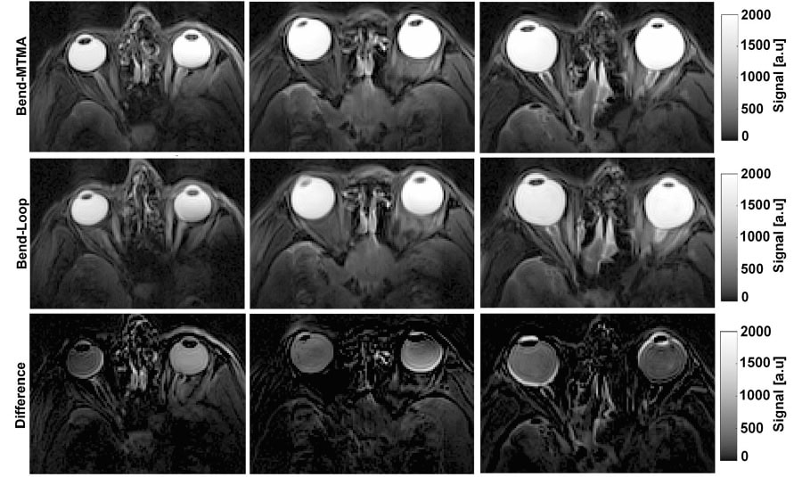
The team first performed MRI of the eye and orbit in three healthy adults, using the bend-loop and bend-MTMA antennas positioned over the eyes. Across all volunteers, the bend-MTMA exhibited better transmit performance in the ocular region that the bend-loop.

The bend-MTMA antenna also generated larger intraocular signals than the bend-loop (assessed via T2-weighted turbo spin-echo imaging), with signal increases of 51%, 28% and 25% in the left eyes, for volunteers 1, 2 and 3, respectively, and corresponding gains of 27%, 26% and 29% for their right eyes. Overall, the bend-MTMA provided more uniform and higher-intensity signal coverage of the ocular region at 7.0 T than the bend-loop.

To further demonstrate clinical application of the bend-MTMA, the team used it to image a volunteer with a retinal haemangioma in their left eye. A 7.0 T MRI scan performed 16 days after treatment revealed two distinct clusters of structural change due to the therapy. In addition, one of the volunteer’s ocular scans revealed a sinus cyst, an unexpected finding that showed the diagnostic benefit of the bend-MTMA being able to image beyond the orbit and into the paranasal sinuses and inferior frontal lobe.

The team used the planar antennas to image the occipital lobe, a clinically relevant target for neuro-ophthalmic examinations. The planar-MTMA exhibited significantly higher transmit efficiency than the planar-loop, as well as higher signal intensity and wider coverage, enhancing the anatomical depiction of posterior brain regions.

“Clearer signals and better images could open new doors in diagnostic imaging,” says Niendorf. “Early ophthalmology applications could include diagnostic confirmation of ambiguous ophthalmoscopic findings, visualization and local staging of ocular masses, 3D MRI, fusion with colour Doppler ultrasound, and physio-metabolic imaging to probe iron concentration or water diffusion in the eye.”

He notes that with slight modifications, the new antennas could enable MRI scans depicting the release and transport of drugs within the body. Their geometry and design could also be tuned to image organs such as the heart, kidneys or brain. “Another pioneering clinical application involves thermal magnetic resonance, which adds a thermal intervention dimension to an MRI device and integrates diagnostic guidance, thermal treatment and therapy monitoring facilitated by metamaterial RF antenna arrays,” he tells Physics World.

The post Metamaterial antennas enhance MR images of the eye and brain appeared first on Physics World.

  •  

Todd McNutt: how an AI software solution enables creation of the best possible radiation treatment plans

Todd McNutt is a radiation oncology physicist at Johns Hopkins University in the US and the co-founder of Oncospace, where he led the development of an artificial intelligence (AI)-powered tool that simultaneously accelerates radiation planning and elevates plan quality and consistency. The software, now rebranded as Plan AI and available from US manufacturer Sun Nuclear, draws upon data from thousands of previous radiotherapy treatments to predict the lowest possible dose to healthy tissues for each new patient. Treatment planners then use this information to define goals that streamline and automate the creation of a best achievable plan.

Physics World’s Tami Freeman spoke with McNutt about the evolution of Oncospace and the benefits that Plan AI brings to radiotherapy patients and cancer treatment centres.

Can you describe how the Oncospace project began?

Back in 2007, several groups were discussing how we could better use clinical data for discovery and knowledge generation. I had several meetings with folks at Johns Hopkins, including Alex Szalay who helped develop the Sloan Digital Sky Survey. He built a large database of galaxies and stars and it became a huge research platform for both amateur and professional astronomers.

From that discussion, and other initiatives, we looked at moving towards structured data collection for patients in the clinical environment. By marrying these data with radiation treatment plans we could study how dose distributions across the anatomy affect patient outcomes. And we took that opportunity to build a database for radiotherapy.

What inspired the transition from academic research to founding the company Oncospace Inc in 2019?

After populating the database with data from many patients, we could examine which anatomic features impact our ability to generate a plan that minimizes radiation dose to normal tissues while treating target volumes as best as possible. We came up with a feature set that characterized the relationships between normal anatomy and targets, as well as target complexity.

This early work allowed us to predict expected doses from these shape-relationship features, and it worked well. At that point, we knew we could tap into this database and generate a prediction that could help create treatment plans for new patients. We thought of this as personalized medicine: for the first time, we could see the level of treatment plan quality that we could achieve for a specific patient.

I thought that this was useful commercially and that we should get it out to other clinics. Praveen Sinha, who I’d known from my previous work at Philips and now leads Sun Nuclear’s software business line, asked if I wanted to create a startup. The timing was right for both of us and I had a team here ready to go, so we went ahead and did it. With his knowledge of startups and my knowledge of what we wanted to achieve, we had perfect timing and a perfect group to work with.

Plan AI enables both predictive planning and peer review, how do these functions work?

The idea behind predictive planning is that, for a given patient, I can predict the expected dose that I should be able to achieve for them.

Plan AI software
Plan AI software Comparing dose–volume histogram prediction bands with clinical goals (arrows) provides users with valuable feedback on what can be achieved before the planning process begins. The screen shows the prediction sent to the treatment planning system.
Dose–volume histograms
Clinical plan The screen shows a review of the results that the treatment planning system achieved, with dose–volume histograms shown by the solid lines.

Treatment planning involves specifying dosimetric objectives to the planning system and asking it to optimize radiation delivery to meet these. But nobody really knows what the right objectives even are – it is just a trial-and-error process. Plan AI’s prediction provides a rational set of objectives for plan optimization, allowing the planning system’s algorithm to move towards a good solution and making treatment planning an easier problem to solve.

Peer review involves a peer physician looking at every treatment plan to evaluate it for quality and safety. But again, people don’t really know the level of quality you can generate, it depends on the patient’s anatomy. Providing a predicted dose with clinical dose goals enables a rapid review to see whether it is a high-quality plan or not.

In the past we looked at simple things like whether a contour is missing slices or contains discontinuities and Plan AI checks for this, but you can do far more with AI. For example, you could look at all the contoured rectums in the system and predict if your contour goes too far into the sigmoid colon, then it may be mis-contoured. We have research software that can flag such potential anomalies so they don’t get overlooked.

The Plan AI models are developed using Oncospace’s database of previous treatments; can you describe this data lake?

When we first started, we developed a large SQL database containing all the shape-relationship features and dosimetry features. The SQL language is ideal for being able to query and sift through the data, but when the company was formed, we recognized that there was some age to that technology.

So for the Plan AI data lake, we extracted all the different shape-relationship and shape-complexity features and put them into a Parquet database in the cloud. This made the data lake much more amenable to applying machine learning algorithms to it. The SQL data lake at Johns Hopkins is maintained separately and primarily used to investigate toxicity predictions and spatial dose patterns. But for Plan AI, the models are fixed and streamlined for the specific task of dose prediction.

What does the model training process entail?

One of the first tasks was to curate the data, using the AAPM’s standardized structure-naming model. Our data scientist Julie Shade wrote some tools for automatic name mapping and target identification; that helped us process much larger amounts of data for the model.

Once we had all the shape-relationship and shape-complexity features and all the doses, we trained the models by anatomical region. We have FDA-approved models for the male and female pelvis, thorax, abdomen and head-and-neck. For each of these, we predict the doses for every organ-at-risk. Then we used a five-fold validation model to make sure that the predictions were good on an internal data set.

We also performed external validation at institutions including Johns Hopkins and Montefiore hospitals. We created predicted plans from recent treatment plans that had been evaluated by physicians. For almost all cases, both plan quality and plan efficiency were improved with Plan AI.

One aspect of this training is that whenever we drive optimization via predictive planning we want to push towards the best achievable dose. Regular machine learning predicts an expected, or average, dose across all patients. But you never want to drive a treatment plan towards the average dose, because then every plan you generate will be happy being average. Our model predicts both the average and the best achievable dose, and drives plan optimization towards the best achievable.

When implementing new technology in the clinic, it’s important to fit into the existing treatment workflow. How clinic-ready are these AI tools?

Radiation therapy is protocol-driven: we know what technique we’re going to use to treat and what our clinical dose goals are for different structures. What we don’t know is the patient-specific part of that. So for each anatomical region, we built models out of a wide range of treatment protocols, with many different types of patients, to ensure that the same prediction model works for any protocol. This means a user can use any protocol for treatment and the predictions will work, they don’t have to retrain anything. It’s ready to go out of the box, there’s a library of protocols to start with, and you can change protocols as you need for your own clinic.

The other part of being clinic-ready is aligning with the way that planning is currently performed, which is using dose–volume histograms. Treatment plans are optimized by manipulating these dose objectives, and that’s exactly what we predict. So users aren’t changing the whole paradigm of how planners operate. They still use their treatment planning system (TPS) – we just put the objectives in there. Basically, a TPS script sends the patient’s CT and contours to the cloud, where Plan AI makes the predictions. The TPS then pulls back in the objectives built from the models, based on this specific patient’s anatomy. The TPS runs the optimization and, as a last step, can send the plan back to Plan AI to check that it fits within the best achievable predictions.

Did you encounter any challenges bringing AI into a clinical setting?

Interestingly, the challenges aren’t technical, they are more human related. One of the more systemic challenges is data security when using medical data for training. A nice thing about our system is that the features we generate from treatment plans are just mathematical shape-relationship features and don’t involve a lot of identifiable information.

AI has been used in radiation therapy for image contouring and auto-segmentation, and early efforts were not so good. So, there’s always a good, healthy scepticism. But once you show people that it works and works well, this can be overcome. I have seen some people worried about job security and AI taking over. We are medical professionals designing a treatment plan to care for a patient and there’s a lot of pride and art in that – if you automate that, it takes away some of this pride and art.

I tell people that if we automate the easier things, then they can spend their quality time on the more difficult and challenging cases, because that’s where their talent might be needed more.

Do you have any advice for clinics looking to adopt AI-driven planning?

Introduce it as an assistant, not as a solution. You want people that already know what they’re doing to be able to use their knowledge more efficiently. We want to make their jobs easier and show them that it also improves quality.

With dosimetrists, for example, they create a plan and work hard getting the dose down – and then the physician looks at it and suggests that they can do better. Predictive planning gives them confidence that they are right and takes the uncertainty out of the physician review process. And once you’ve gained that level of confidence, you can start using it for adaptive planning or other technologies.

Where do you see predictive modelling and AI in oncology in five years from now?

Right now, there’s been a lot of data collected, but we want that data to advance and learn. Having multiple centres adding to this pool of knowledge and being able to continually update those models from new, broader data sets could be of huge value.

In terms of patient outcomes, we’ve done a lot of the work looking at how the spatial pattern of dose impacts toxicity and outcomes. This is part of the research being performed at Johns Hopkins and still in discovery mode. But down the road, some of these predictions of normal tissue outcomes could be fed into the planning process to help reduce toxicity at the patient level.

Finally, what’s been the most rewarding part of this journey for you?

During my prior experience building treatment planning systems, the biggest problem was always that nobody knew what the objective was. Nobody knew how to tell the system: “this is the dose I expect to receive, now optimize to get it for me”, because you didn’t know what you could do. For any given patient, you could ask for too much or too little. Now, for the first time, I argue that we actually know what our objective is in our treatment planning.

This levels the playing field between different environments, different countries, or even different dosimetrists with different levels of experience. The Plan AI tool brings all this to a consistent state and enables high quality, efficient planning everywhere. We can provide this predictive planning tool to clinics around the world. Now we just have to get everybody using it.


The post Todd McNutt: how an AI software solution enables creation of the best possible radiation treatment plans appeared first on Physics World.

  •  

New journal aims to advance the interdisciplinary field of personalized health

Personalized health – the use of individualized measurements to address each patient’s specific needs – is a research field that’s evolving at pace. Bringing this level of personalization into the clinic is an interdisciplinary challenge, requiring the development of sensors that generate clinically meaningful data outside the hospital, new imaging modalities and analysis techniques, and computational tools that address the uncertainties of dealing with just one individual.

Much of the most impactful work in this field sits in the spaces between established disciplines. And for researchers looking to publish their findings or read about the latest breakthroughs, this work is often scattered across discipline-specific journals. A new open access journal from IOP Publishing – Medical Sensors & Imaging (MSI) – aims to remedy this shortfall, providing a dedicated home for authors working across sensing, imaging, modelling and data-driven healthcare.

Medical Sensors & Imaging
New launch Medical Sensors & Imaging is fully open for submissions. (Courtesy: IOP Publishing)

“We want a journal where physicists, engineers, computer scientists, biomedical researchers and clinicians can publish and read work that advances personalized health, without confinement into traditional silos,” explains founding editor-in-chief Marco Palombo from Cardiff University. “MSI also aims to play an important role in strengthening interdisciplinary exchange.”

“The community needs a specialized forum that doesn’t just report on new materials or a clinical trial, but validates innovations that can specifically solve complex biomedical challenges,” adds deputy editor Xiliang Luo from Qingdao University of Science and Technology. “I think this journal is a perfect fit for that gap.”

Connecting communities

Published by IOP Publishing on behalf of the Institute of Physics and Engineering in Medicine (IPEM), MSI aims to dismantle the barriers between engineering innovation and clinical application by creating a community of experts that work together to translate innovative technology into clinical settings.

MSI sits within IPEM’s journal portfolio that includes Physics in Medicine & Biology, Physiological Measurement and Medical Engineering & Physics. Its aims and scope were designed to complement, rather than overlap with, these existing journals and provide a dedicated venue for translational work and practical applied research that may otherwise struggle to fit a traditional scope.

Marco Palombo
Marco Palombo: “We have been discussing green AI and green healthcare for at least 10 years. I think MSI can be one of the first journals to push this area forward.”

Being part of this established family of journals brings with it strong editorial standards, an established readership base and a commitment to scientific integrity. The journal also offers rapid, high-quality peer review, with feedback that’s constructive, rigorous and fair. MSI is fully open access, which maximizes the visibility, reach and impact of its published papers.

“For a new journal in a dynamic field, ensuring content is discoverable and barrier-free is essential for building an audience quickly and establishing credibility,” says Palombo. “We also wanted MSI to support global participation. Many excellent groups operate with limited budgets but make major scientific contributions. Open access reduces inequities in who can read and build on published work.”

“For the authors, we can provide a specialized platform for scientists whose work transcends traditional boundaries, offering visibility to a broad audience that’s eager for translational solutions,” says Luo. “And for the readers, I think we will be the go-to resource for academic researchers, industry R&D leaders, and healthcare innovators seeking the latest breakthroughs in personalized health monitoring and advanced diagnostics.”

Hot topics

Palombo contributed to the strategic development of the journal at an early stage, drawing upon his experience in healthcare and medical imaging research and engaging with the research community to identify the scientific niche that MSI could fill. Working with IOP Publishing, he helped shape the journal’s aims and scope and assembled a diverse, internationally recognized editorial board with knowledge aligned with the journal’s mission – including Lui, who brings specialist expertise in wearable technologies and biosensors.

Xiliang Luo
Xiliang Luo: “We hope to establish a forum where advanced sensing technology and imaging techniques can enable the next generation of personalized and predictive health.”

The journal will publish high-quality research on novel biomedical sensing and imaging techniques, along with the algorithms, validation frameworks and translational studies that demonstrate their application in real-world medicine. MSI also provides a platform to showcase research on hot topics such as wearable and implantable sensors for continuous physiological monitoring, for example, or microneedle-based sensing technologies and breath analysis.

The development of flexible and biocompatible materials will be key for the growth of bio-integrated devices and biodegradable or transient electronics, as will anti-fouling strategies that enable use of sensors in complex biological environments. On the imaging side, the journal scope encompasses mainstay medical imaging techniques such as MRI, CT, ultrasound, PET and SPECT, as well as emerging multimodal and hybrid approaches, with a focus on technical innovation and translational relevance.

“Given my own background, I’m particularly keen to see strong submissions in the area of MRI, including advanced quantitative biomarkers and approaches that probe tissue microstructure,” notes Palombo. “I also see huge potential in connecting imaging to computational modelling – particularly digital twins – and in building imaging pipelines that enable personalized diagnosis and prognosis.”

“Other exciting areas include combining sensing and imaging technologies into one system, and closed-loop ‘sense then act’ systems, which sense something and can then release medicine to treat the disease,” says Luo.

The rise of AI

Artificial intelligence (AI) is becoming increasingly central to both sensing and imaging, and will likely play a major role in the evolution of personalized health, enabling a shift towards multimodal fusion of sensor streams, imaging and clinical data. AI could also facilitate the introduction of integrated sensor systems that collect data and interpret signals in real time, and digital twins that link patient-specific data with computational models to simulate disease progression or treatment response.

Palombo emphasizes the importance of trustworthy AI: methods that don’t just provide an output, but are explainable, robust and explicitly handle uncertainty. This is a direction seen in the general field of AI, but is especially important within healthcare. He also cites the increasing momentum around green healthcare and green AI, with personalized health technologies designed to reduce waste and minimize energy consumption, and clinical models developed with far greater computational efficiency.

“It would be fantastic to have an AI model running directly on the sensor, for example, and this ties in with the environmental impact of AI,” he explains. “If we keep AI small and manageable, then it pollutes less, is more affordable for everybody and can be deployed on small, lightweight devices.”

A community focal point

Looking ahead, Palombo hopes that MSI will becomes a leading platform for interdisciplinary innovation in personalized health, and the routine home for publishing major advances in sensing, imaging, modelling and trustworthy AI. “Over time, I’d like the journal to build depth in core areas, while also actively shaping emerging directions such as digital twins, uncertainty-aware and explainable AI, multimodal integration and technologies that are genuinely deployable in clinical workflows.”

“Currently, the fields of sensor engineering and clinical medicine often run on parallel tracks. My hope is that this journal will force these tracks to converge over time,” adds Luo. “I see the journal fostering a new language where chemists, physicists, engineers and doctors can understand each other by publishing papers in MSI.”

  • Medical Sensors & Imaging is fully open for submissions, with the first issue expected to publish in Q2/Q3 of this year. During the launch phase, IOPP is covering the article processing charge (APC) for all accepted papers, enabling early contributors to publish at no cost while helping the journal establish a strong foundation of high-quality inaugural content. Beyond this period, many authors will benefit from support through IOPP’s transformative agreements, while others may be eligible for APC waivers and discounts.


The post New journal aims to advance the interdisciplinary field of personalized health appeared first on Physics World.

  •  

World’s smallest QR code paves the way for ultralong-life data storage

A team headed up at TU Wien in Austria has set the Guinness World Record for creating the world’s smallest QR code. Working with industry partner Cerabyte, the researchers produced a stable and repeatedly readable QR code with an area of just 1.977 µm2. When read out – using an electron microscope, as its structure is too fine to be seen with a standard optical microscope – the QR code links to a scientific webpage at TU Wien.

But this wasn’t just a ploy to get into the record books, the QR code was created as part of the team’s research into ceramic data storage materials. Unlike conventional magnetic or electronic data storage media, which degrade within decades, ceramic-based storage is designed to withstand extreme temperatures, radiation, chemical corrosion and mechanical damage.

As such, information stored in ceramic materials could endure for centuries, or even millennia. And in contrast to today’s data centres, ceramics preserve stored information without any energy input and without requiring cooling.

Electron microscope image of QR code
Invisible code The world’s smallest QR code can only be read out using an electron microscope. (Courtesy: TU Wien)

To create these ultralong-life data storage systems, the researchers use focused ion beams to mill the QR code into a thin film of chromium nitride, a durable ceramic often used to coat high-performance cutting tools. As each individual pixel is just 49 nm in size, roughly 10 times smaller than the wavelength of visible light, the code cannot be imaged using visible light. But when examined with an electron microscope, the QR code could indeed be read out reliably.

After the writing process, the entire stack of ceramic films is subjected to extreme conditions, such as high temperatures, corrosive environments and mechanical stress, to evaluate the material’s long-term durability and readout stability.

Pushing storage to its limits

Creating a “tiny QR code” was not the team’s initial goal, but emerged as a natural outcome of pushing this storage technology to its limits, says Paul Mayrhofer from TU Wien’s Institute of Materials Science and Technology.

“During a discussion with one of my PhD students, Erwin Peck, we realised that the writing procedure we had developed already produced features smaller than what had previously been reported for QR codes,” he explains. “This sparked the idea: if we can reliably write structures at that scale, why not intentionally create the smallest QR code possible?”

To claim its place in the record books, the QR code was successfully milled and read out in the presence of witnesses and its size independently verified using calibrated scanning electron microscopy at the University of Vienna. It is now officially recognized by Guinness as the world’s smallest QR code, and is roughly one third the size of the previous record holder.

Mayrhofer points out that the storage capacity of the ceramic data storage technology far surpasses that of a single QR code. “Based on current estimates, a cartridge of 100 x 100 x 20 mm with ceramic storage medium could potentially store on the order of 290 terabytes of raw data,” he says.

As well as offering this impressive raw capacity, for practical applications it’s also crucial that the ceramic storage offers high writing speed, which determines how efficiently large datasets can be stored, and low energy consumption during writing, which will influence the potential for scalability and sustainability. The researchers are currently working to optimize both of these parameters.

“Humanity has preserved information for millennia when carved in stone, yet much of today’s digital information risks being lost within decades,” project leader Alexander Kirnbauer tells Physics World. “Our long-term goal is to create an ultrastable, sustainable data storage technology capable of preserving information for extremely long times – potentially thousands to millions of years. In essence, we want to develop a form of storage that ensures the knowledge of our digital age does not disappear over time.”

The post World’s smallest QR code paves the way for ultralong-life data storage appeared first on Physics World.

  •  

Nickel-enhanced biomaterial becomes stronger when wet

Synthetic materials such as plastics are designed to be durable and water resistant. But the processing required to achieve these properties results in a lack of biodegradability, leading to an accumulation of plastic pollution that affects both the environment and human health. Researchers at the Institute for Bioengineering of Catalonia (IBEC) are developing a possible replacement for plastics: a novel biomaterial based on chitin, the second most abundant natural polymer on Earth.

“Every year, nature produces on the order of 1011 tonnes of chitin, roughly equivalent to more than three centuries of today’s global plastic production,” says study leader Javier G Fernández. “Chitin and [its derivative] chitosan are the ultimate natural engineering polymers. In nature, variations of this material produce stiff insect wings enabling flight, elastic joints enabling extraordinary jumping in grasshoppers, and armour-like protective exoskeletons in lobsters or clams.”

But while biomaterials provide a more environmentally friendly alternative to conventional plastics, most biological materials weaken when exposed to water. In this latest work, Fernández and first author Akshayakumar Kompa took inspiration from nature and developed a new biomaterial that increases its strength when in contact with water, while maintaining its natural biodegradability.

Metal matters

In the exoskeletons of insects and crustaceans, chitin it is secreted in a gel-like form into water and then transitions into a hard structure. Following a chance observation that removing zinc from a sandworm’s fangs caused them to soften in water, Kompa and Fernández investigated whether adding a different transition metal, nickel, to chitosan could have the opposite effect.

By mixing nickel chloride solution (at concentrations from 0.6 to 1.4 M) with dispersions of chitosan extracted from discarded shrimp shells, the researchers entrapped varying amounts of nickel within the chitosan structure. Fourier-transform infrared spectra of resulting chitosan films revealed the presence of nickel ions, which form weak hydrogen bonds with water molecules and increase the biomaterial’s capacity to bond with water.

“In our films, water molecules form reversible bridges between polymer chains through weak interactions that can rapidly break and reform under load,” Fernández explains. “That fast reconfiguration is what gives the material high strength and toughness under wet conditions: essentially a built-in, stress-activated ‘self-rearrangement’ mechanism. Nickel ions act as stabilizing anchors for these water-mediated bridges, enabling more and longer-range connections and making inter-chain connectivity more robust”.

The nickel-doped chitosan samples had tensile strengths of between 30 and 40 MPa, similar to that of standard plastics. Adding low concentrations of nickel did not significantly impact the mechanical properties of the films. Concentrations of 1 M or more, however, preserved the material’s strength while increasing its toughness (the ability to stretch before breaking) – a key goal in the field of structural materials and a feature unique to biological composites.

Testing a nickel-doped chitosan film
Increased strength Testing a nickel-doped chitosan film using a 20 kg dumbbell. (Courtesy: Institute for Bioengineering of Catalonia)

Upon immersion in water, the nickel-doped films exhibited greater tensile strength, increasing from 36.12±2.21 MPa when dry to 53.01±1.68 MPa, moving into the range of higher-performance engineering plastics. In particular, samples created from an optimal 0.8 M nickel concentration almost doubled in strength when wet (and were used for the remainder of the team’s experiments).

Scaling production

The manufacturing process involves an initial immersion in water, followed by drying for 24 h and then re-wetting. During the first immersion, any nickel ions that are not incorporated into the material’s functional bridging network are released into the water, ensuring that nickel is present only where it is structurally useful.

The researchers developed a zero-waste production cycle in which this water is used as a primary component for fabricating the next object. “The expelled nickel is recovered and used to make the next batch of material, so the process operates at essentially 100% nickel utilization across batches,” says Fernández.

Nickel-doped chitosan structures
Zero waste production The team created structures including a 3 m2 nickel-doped chitosan film and a cup that can retain water as effectively as common plastics. (Courtesy: Institute for Bioengineering of Catalonia)

They used this process to produce various nickel-doped chitosan objects, including watertight containers and a 1 m2 film that could support a 20 kg weight after 24 h of water immersion. They also created a 244 x 122 cm film with similar mechanical behaviour to the smaller samples, demonstrating the potential for rapid scaling to ecologically relevant scales. A standard half-life test revealed that after approximately four months buried in garden soil, half of the material had biodegraded.

The researchers suggest that the biomaterial’s first real-world use may be in sectors such as agriculture and fishing that require strong, water-compatible and ultimately biodegradable materials, likely for packaging, coatings and other water-exposed applications. Both nickel and chitosan are already employed within biomedicine, making medicine another possible target, although any new medical product will require additional regulatory and performance validation.

The team is currently setting up a 1000 m2 lab facility in Barcelona, scheduled to open in 2028, for academia–industry collaborations in sustainable bioengineering research. Fernández suggests that we are moving towards a “biomaterial age”, defined by the ability to “control, integrate, and broadly use biomaterials and biological principles within engineering applications”.

“Over the last 20 years, working on bioinspired manufacturing, we have been able to produce the largest bioprinted objects in the world, demonstrated pathways for resource-secure and sustainable production in urban environments, and even explored how these approaches can support interplanetary colonization,” he tells Physics World. “Now we are achieving material properties that were considered out of reach by designing the material to work with its environment, rather than isolating itself from it.”

The researchers report their findings in Nature Communications.

The post Nickel-enhanced biomaterial becomes stronger when wet appeared first on Physics World.

  •  

Dual-tracer PET enables biologically individualized radiotherapy

Radiation therapy is usually delivered by prescribing the same radiation dose for each particular type of tumour. But this “one-size-fits-all” approach does not account for a tumour’s intrinsic radiosensitivity and heterogeneity and can lead to recurrence and treatment failure. Researchers in Sweden and Germany are now investigating whether biologically individualized radiotherapy plans, created using PET images of a patient’s tumour biology, can improve treatment outcomes.

The research team – headed up by Marta Lazzeroni from Stockholm University – studied 28 patients with advanced head-and-neck squamous cell carcinoma (HNSCC). All patients underwent two pre-treatment PET/CT scans, using 18F-fluoromisonidazole (FMISO) and 18F-FDG as tracers to respectively quantify radioresistance and tumour cellularity (the percentage of clonogenic cells) – both critical factors that influence treatment response.

“FMISO provides information on hypoxia-related radioresistance, but tumour control also strongly depends on the number of clonogenic cells, which is not captured by hypoxia imaging alone,” Lazzeroni explains. “To our knowledge, this is the first study to combine FMISO and FDG PET within a unified radiobiological framework to guide biologically individualized dose escalation.”

For each patient, the researchers used FMISO uptake to derive voxel-level maps of oxygen partial pressure (pO2) in the tumour and define a hypoxic target volume (HTV). The FDG scans were used to estimate spatial variations in clonogenic tumour cell density, which directly influence the dose required to realise a given tumour control probability (TCP).

Based on individual tumour profiles, the team used automated planning to create volumetric-modulated arc therapy plans comprising 35 fractions with an integrated boost. The plans delivered escalated dose to radioresistant subvolumes (the HTV), while maintaining clinically acceptable sparing of organs-at-risk. The PET datasets were used to calculate the prescribed dose required to achieve a TCP of 95%.

Meeting clinical feasibility

The automated planning pipeline achieved high-quality treatment plans for all patients without manual intervention. The average EQD2 (the dose delivered in 2 Gy fractions that’s biologically equivalent to the total dose) to the HTV was boosted to 81±3.2 Gy, and all 28 plans met the clinical constraints for protecting the brainstem, spinal cord and mandible. Parotid glands were spared in 75% of cases, with the remainder being glands that overlapped the target.

Lazzeroni and colleagues suggest that these results confirm the overall clinical feasibility of their personalized dose-escalation strategy and demonstrate how biology-guided prescriptions could be integrated into existing treatment planning workflows.

The researchers also performed a radiobiologic evaluation of the treatment plans to see whether the optimized dose distribution achieved the desired target control. For this, they calculated the TCP based on the planned dose distribution, the PET-derived radioresistance data and clonogenic cell density maps. For all patients, the plans achieved model-predicted TCP values exceeding 90%, a notable improvement on tumour control rates reported in the clinical literature for HNSCC, which are typically around 60%.

The proposed strategy is based on pre-treatment PET images, but biological changes during treatment – including temporal and spatial variations in tumour hypoxia – could impact its effectiveness. In future, the researchers suggest that longitudinal imaging, such as PET/CT scans at weeks 3 and 5, could be used to monitor evolving tumour biology and inform adaptive replanning. This is particularly relevant in HNSCC, where tumour shrinkage and reoxygenation are common, and where updated imaging is required to determine whether dose escalation or de-escalation is appropriate to maintain tumour control and optimize normal tissue sparing.

The researchers point out that as the biology-guided dose prescriptions were planned but not delivered, prospective trials will be required to assess whether the observed dosimetric and biologic gains translate to improved patient outcomes.

“This study was designed as a feasibility and modelling investigation, and the next step is prospective clinical validation,” Lazzeroni tells Physics World. “Based on the promising results of this approach, prospective clinical trials are currently in the planning phase within the group led by Anca-L Grosu in Germany. These trials will focus on integrating longitudinal PET imaging during treatment to enable biologically adaptive radiotherapy.”

The results are published in the Journal of Nuclear Medicine.

The post Dual-tracer PET enables biologically individualized radiotherapy appeared first on Physics World.

  •  

Implanted electrodes provide intuitive control of prosthetic hand

Loss of a limb can significantly impact a person’s independence and quality-of-life, with arm amputations particularly impeding routine daily activities. Prosthetic limbs can restore some of the lost function, but often rely on surface electrodes with low signal quality. A research team at the University of Michigan has now shown that implanted electrodes could provide more accurate and reliable control of hand and wrist prostheses.

Today, most upper-limb prostheses are controlled using surface electrodes placed on the skin to detect electrical activity from underlying muscles. The recorded electromyography (EMG) signals are then used to classify different finger and wrist movements. Under real-world conditions, however, these signals can be impaired by inconsistent electrode positioning, changes in limb volume, exposure to sweat and artefacts from user movements.

Implanted electrodes, tiny contacts that are surgically sutured into muscles, could do a better job. By targeting muscles deeper in the arm, they offer higher signal-to-noise ratios and less susceptibility to daily variations. And although amputation can eliminate many of the muscles that control hand functions, techniques such as regenerative peripheral nerve interface (RPNI) surgery – in which muscle tissue is grafted to nerves in the residual limb – enable electrodes to target missing muscles and record relevant signals for prosthetic control.

Senior author Cynthia Chestek points out that such RPNI grafts are also beneficial for the nerve itself. “They provide a target for nerve endings that prevent the formation of painful neuromas, and that may in turn help reduce phantom limb pain,” she explains “In future, it would also be possible to place electrodes and a wireless transmitter during that same surgery, such that no additional surgeries are required other than the original amputation.”

In their latest work, reported in the Journal of Neural Engineering, Chestek and colleagues investigated whether implanted electrodes could provide stable and high-quality signals for  controlling prosthetic hand and wrist function.

Performance comparisons

The study involved two individuals with forearm amputations and EMG electrodes implanted into RPNIs and muscles in their residual limb. The subjects performed various experiments, during which the team recorded EMG signals from the implanted electrodes plus dry-domed and gelled (used to improve contact with the skin) surface electrodes.

In one experiment, participants were tasked with controlling a virtual hand and wrist in real time by mimicking movements (various grips) on a screen. The researchers used the recorded EMG signals to train linear discriminant analysis classifiers to distinguish the cued grips, training separate classifiers for each electrode type.

They then evaluated the performance of these grip classifiers during a posture classification experiment, in which the subjects actively controlled hand or wrist movements of a virtual hand. Participants achieved faster, more accurate and more reliable control using the implanted electrodes than the surface electrodes.

With participants sitting and keeping their arm still, the implanted electrodes achieved average per-bin accuracies (the percentage of correctly classified time bins) of 82.1% and 91.2% for subjects 1 and 2, respectively. The surface electrodes performed worse, with accuracies of 77.1% and 81.3% for gelled electrodes, and 58.2% and 67.1% for dry-domed electrodes, for subjects 1 and 2, respectively.

The researchers repeated this experiment with the subjects standing and moving their arm to mimic daily activities. Adding movement reduced the classification accuracy in all cases, but affected the implanted electrodes to a far smaller degree. The control success rate (the ability to hold a grip for at least 1 s, within 3 s of seeing a movement cue) also diminished between still and moving conditions, but again, the implanted electrodes experienced smaller decreases.

Overall, the performance of online classifiers using implanted electrodes was only slightly affected by arm movements, while classifiers trained on surface electrodes became unstable. Investigating the reasons underlying this difference revealed that implanted electrodes exhibited higher EMG signal amplitudes, lower cross-correlation between channels, and smaller signal deviations between still and moving conditions.

The Coffee Task

To examine a real-world scenario, subject 1 completed the “Coffee Task”, which involves performing the various grips and movements required to: place a cup into a coffee machine; place a coffee pod into the machine; push the start button; move the filled cup onto a table; and open a sugar packet and pour it into the cup.

The subject performed the task using an iLimb Quantum myoelectric prosthetic hand controlled by either implanted or dry surface electrodes, with and without control of wrist rotation. The participant performed the task faster using implanted electrodes, successfully completing the task on all three attempts. For surface-based control, they reached the maximum time limit of 150 s in two out of three attempts.

Although gelled electrodes are the gold standard for surface EMG, they cannot be used whilst wearing a standard prosthetic socket. “With the Coffee Task, use of the physical prosthetic  hand is needed, so this was only performed with dry-domed surface electrodes and implanted electrodes,” explains first author Dylan Wallace.

The researchers also assessed whether simultaneous wrist and hand control can reduce compensatory body movements (measured using reflective markers on the subject’s torso), compared with hand control alone. Without wrist rotation, the subject had to lean their entire upper body to complete the pouring task. With wrist rotation enabled, this lean was greatly reduced.

This finding emphasizes how wrist control provides significant functional benefit for prosthesis users during daily activities. Chestek notes that in a previous study where participants wore a prosthesis without an active wrist, “almost everything we asked them to do required large body movements”.

“Fortunately, the implantable electrodes provide highly specific and high-amplitude signals, such that we were able to add that wrist movement without losing the ability to classify multiple different grasps,” she explains. “The next step would be to pursue continuous, rather than discrete, movement for all of the individual joints of the hand –  though that will not happen quickly.”

The post Implanted electrodes provide intuitive control of prosthetic hand appeared first on Physics World.

  •  

Multi-ion cancer therapy tackles the LET trilemma

Cancer treatments using heavy ions offer several key advantages over conventional proton therapy: a sharper Bragg peak and small lateral scattering for precision tumour targeting, as well as high linear energy transfer (LET). High-LET radiation induces complex DNA damage in cancer cells, enabling effective treatment of even hypoxic, radioresistant tumours. A team at the National Institutes for Quantum Science and Technology (QST) in Japan is now exploring the potential benefits of multi-ion therapy combining beams of carbon, oxygen and neon ions.

“Different ions exhibit distinct physical and biological characteristics,” explains QST researcher Takamitsu Masuda. “Combining them in a way that is tailored to the specific characteristics of a tumour and its environment allows us to enhance tumour control while reducing damage to surrounding healthy tissues.”

The researchers are using multi-ion therapy to increase the dose-averaged LET (LETd) within the tumour, performing a phase I trial at the QST Hospital to evaluate the safety and feasibility of this LETd escalation for head-and-neck cancers. But while high LETd prescriptions can improve treatment efficacy, increasing LETd can also deteriorate plan robustness. This so-called “LET trilemma” – a complex trade-off between target dose homogeneity, range robustness and high LETd – is a major challenge in particle therapy optimization.

In their latest study, reported in Physics in Medicine & Biology, Masuda and colleagues evaluated the impact of range and setup uncertainties on LETd-optimized multi-ion treatment plans, examining strategies that could potentially overcome this LET trilemma.

Robustness evaluation

The team retrospectively analysed the data of six patients who had previously been treated with carbon-ion therapy. Patients 1, 2 and 3 had small, medium and large central tumours, respectively, and adjacent dose-limiting organs-at-risk (OARs); and patients 4, 5 and 6 had small, medium and large peripheral tumours and no dose-limiting OARs.

Multi-ion therapy plans
Multi-ion therapy plans Reference dose and LETd distributions for patients 1, 2 and 3 for multi-ion therapy with a target LETd of 90 keV/µm. The GTV, clinical target volume (CTV) and OARs are shown in cyan, green and magenta, respectively. (Courtesy: Phys. Med. Biol.10.1088/1361-6560/ae387b)

For each case, the researchers first generated baseline carbon-ion therapy plans and then incorporated oxygen- or neon-ion beams and tuned the plans to achieve a target LETd of 90 keV/µm to the gross tumour volume (GTV).

Particle therapy plans can be affected by both range uncertainties and setup variations. To assess the impact of these uncertainties, the researchers recalculated the multi-ion plans to incorporate range deviations of +2.5% (overshoot) and –2.5% (undershoot) and various setup uncertainties, evaluating their combined effects on dose and LETd distributions.

They found that range uncertainty was the main contributor to degraded plan quality. In general, range overshoot increased dose to the target, while undershoot decreased dose. Range uncertainties had the largest effect on small tumours and central tumours: patient #1 exhibited a deviation of around ±6% from the reference, while patient #3 showed a dose deviation of just ±1%. Robust target coverage was maintained in all large or peripheral tumours, but deteriorated in patient 1, leading to an uncertainty band of roughly 11%.

“Wide uncertainty bands indicate a higher risk that the intended dose may not be accurately delivered,” Masuda explains. “In particular, a pronounced lower band for the GTV suggests the potential for cold spots within the tumour, which could compromise local tumour control.”

The team also observed that range undershoot increased LETd and overshoot decreased it, although absolute differences in LETd within the entire target were small. Importantly, all OAR dose constraints were satisfied even in the largest error scenarios, with uncertainty bands comparable to those of conventional carbon-ion treatment plans.

Addressing the LET trilemma

To investigate strategies to improve plan robustness, the researchers created five new plans for patient 1, who had a small, central tumour that was particularly susceptible to uncertainties. They modified the original multi-ion plan (carbon- and oxygen-ion beams delivered at 70° and 290°) in five ways: expanding the target; altering the beam angles to orthogonal or opposing arrangements; increasing the number of irradiation fields to a four-field arrangement; and using oxygen ions for both beam ports (“heavier-ion selection”).

The heavier-ion selection plan proved the most effective in mitigating the effects of range uncertainty, substantially narrowing the dose uncertainty bands compared with the original plan. The team attribute this to the inherently higher LETd in heavier ions, making the 90 keV/µm target easier to achieve with oxygen-ion beams alone. The other plan modifications led to limited improvements.

Dose–volume histograms
Improving robustness Dose–volume histograms for patient 1, for the original multi-ion plan and the heavier-ion selection plan, showing the combined effects of range and setup uncertainties. Solid, dashed and dotted curves represent the reference plans, and upper and lower uncertainty scenarios, respectively. (Courtesy: Phys. Med. Biol.10.1088/1361-6560/ae387b)

These findings suggest that strategically employing heavier ions to enhance plan robustness could help control the balance among range robustness, uniform dose and high LETd – potentially offering a practical strategy to overcome the LET trilemma.

“Clinically, this strategy is particularly well-suited for small, deep-seated tumours and complex, variable sites such as the nasal cavity, where range uncertainties are amplified by depth, steep dose gradients and daily anatomical changes,” says Masuda. “In such cases, the use of heavier ions enables robust dose delivery with high LETd.”

The researchers are now exploring the integration of emerging technologies – such as robust optimization, arc therapy, dual-energy CT, in-beam PET and online adaptation – to minimize uncertainties. “This integration is highly desirable for applying multi-ion therapy to challenging cases such as pancreatic cancer, where uncertainties are inherently large, or hypofractionated treatments, where even a single error can have a significant impact,” Masuda tells Physics World.

The post Multi-ion cancer therapy tackles the LET trilemma appeared first on Physics World.

  •  

Polarization-sensitive photoacoustic microscopy reveals heart tissue health

MIR-DS-PAM images of fibrotic and normal cardiac tissue
Imaging tissue fibrosis (a) Mid-infrared dichroism-sensitive photoacoustic microscopy (MIR-DS-PAM) images of cell-induced fibrosis (CIF) and normal control (NC) tissue; (c) MIR-DS-PAM images of drug-induced fibrosis (DIF) and NC tissue; (b) and (d) show the corresponding confocal fluorescence microscopy (CFM) images. Scale bars: 500 µm. (Courtesy: CC-BY 4.0/Light Sci. Appl. 10.1038/s41377-025-02117-0)

Many of the tissues in the human body rely upon highly organized microstructures to function effectively. If the collagen fibres in heart muscle become disordered, for instance, this can lead to or reflect disorders such as fibrosis and cancer. To image and analyse such structural changes, researchers at Pohang University of Science and Technology (POSTECH) in Korea have developed a new label-free microscopy technique and demonstrated its use in engineered heart tissue.

The ability to assess the alignment of microstructures such as protein fibres within tissue’s extracellular matrix provides a valuable tool for diagnosing disease, monitoring therapy response and evaluating tissue engineering models. Currently, however, this is achieved using histological imaging methods based on immunofluorescent staining, which can be labour-intensive and sensitive to the imaging conditions and antibodies used.

Instead, a team headed up by Chulhong Kim and Jinah Jang is investigating photoacoustic microscopy (PAM), a label-free imaging modality that relies on light absorption by endogenous tissue chromophores to reveal structural and functional information. In particular, PAM with mid-infrared (MIR) incident light provides bond-selective, high-contrast imaging of proteins, lipids and carbohydrates. The researchers also incorporated dichroism-sensitive (DS) functionality, resulting in a technique referred to as MIR-DS-PAM.

“Dichroism-sensitivity enables the quantitative assessment of fibre alignment by detecting the polarization-dependent absorption of anisotropic materials like collagen,” explains first author Eunwoo Park. “This adds a new contrast mechanism to conventional photoacoustic imaging, allowing simultaneous visualization of molecular content and microstructural organization without any labelling.”

Park and colleagues constructed a MIR-DS-PAM system using a pulsed quantum cascade laser as the light source. They tuned the laser to a centre wavelength of 6.0 µm to correspond with an absorption peak from the C=O stretching vibration in proteins. The laser beam was linearly polarized, modulated by a half-wave plate and used to illuminate the target tissue.

Tissue analysis

To validate the functionality of their MIR-DS-PAM technique, the researchers used it to image a formalin-fixed section of engineered heart tissue (EHT). They obtained images at four incident angles and used the acquired photoacoustic data to calculate the photoacoustic amplitude, which visualizes the protein content, as well as the degree of linear dichroism (DoLD) and the orientation angle of linear dichroism (AoLD), which reveal the extracellular matrix alignment.

“Cardiac tissue features highly aligned extracellular matrix with complex fibre orientation and layered architecture, which are critical to its mechanical and electrical function,” Park explains. “These properties make it an ideal model for demonstrating the ability of MIR-DS-PAM to detect physiologically relevant histostructural and fibrosis-related changes.”

The researchers also used MIR-DS-PAM to quantify the structural integrity of EHT during development, using specimens cultured for one to five days before fixing. Analysis of the label-free images revealed that as the tissue matured, the DoLD gradually increased, while the standard deviation of the AoLD decreased – indicating increased protein accumulation with more uniform fibre alignment over time. They note that these results agree with those from immunofluorescence-stained confocal fluorescence microscopy.

Next, they examined diseased EHT with two types of fibrosis: cell-induced fibrosis (CIF) and drug-induced fibrosis (DIF). In the CIF sample, the average photoacoustic amplitude and AoLD uniformity were both lower than found in normal EHT, indicating reduced protein density and disrupted fibre alignment. DIF exhibited a higher photoacoustic amplitude and lower AoLD uniformity than normal EHT, suggesting extensive extracellular matrix accumulation with disorganized orientation.

Both CIF and DIF showed a slight reduction in DoLD, again signifying a disorganized tissue structure, a common hallmark of fibrosis. The two fibrosis types, however, exhibited diverse biochemical profiles and different levels of mechanical dysfunction. The findings demonstrate the ability of MIR-DS-PAM to distinguish diseased from healthy tissue and identify different types of fibrosis. The researchers also imaged a tissue assembly containing both normal and fibrotic EHT to show that MIR-DS-PAM can capture features in a composite sample.

They conclude that MIR-DS-PAM enables label-free monitoring of both tissue development and fibrotic remodelling. As such, the technique shows potential for use within tissue engineering research, as well as providing a diagnostic tool for assessing tissue fibrosis or remodelling in biopsied samples. “Its ability to visualize both biochemical composition and structural alignment could aid in identifying pathological changes in cardiological, musculoskeletal or ocular tissues,” says Park.

“We are currently expanding the application of MIR-DS-PAM to disease contexts where extracellular matrix remodelling plays a central role,” he adds. “Our goal is to identify label-free histological biomarkers that capture both molecular and structural signatures of fibrosis and degeneration, enabling multiparametric analysis in pathological conditions.”


The post Polarization-sensitive photoacoustic microscopy reveals heart tissue health appeared first on Physics World.

  •  

RFID-tagged drug capsule lets doctors know when it has been swallowed

Taking medication as and when prescribed is crucial for it to have the desired effect. But nearly half of people with chronic conditions don’t adhere to their medication regimes, a serious problem that leads to preventable deaths, drug resistance and increased healthcare costs. So how can medical professionals ensure that patients are taking their medicine as prescribed?

A team at Massachusetts Institute of Technology (MIT) has come up with a solution: a drug capsule containing an RFID tag that uses radiofrequency (RF) signals to communicate that it has been swallowed, and then bioresorbs into the body.

“Medication non-adherence remains a major cause of preventable morbidity and cost, but existing ingestible tracking systems rely on non-degradable electronics,” explains project leader Giovanni Traverso. “Our motivation was to create a passive, battery-free adherence sensor that confirms ingestion while fully biodegrading, avoiding long-term safety and environmental concerns associated with persistent electronic devices.”

The device – named SAFARI (smart adherence via Faraday cage and resorbable ingestible) – incorporates an RFID tag with a zinc foil RF antenna and an RF chip, as well as the drug payload, inside an ingestible gelatin capsule. The capsule is coated with a mixture of cellulose and molybdenum particles, which blocks the transit of any RF signals.

SAFARI capsules with and without RF-blocking coating
SAFARI capsules Photos of the capsules with (left) and without (right) the RF-blocking coating. (Courtesy: Mehmet Say)

Once swallowed, however, this shielding layer breaks down in the stomach. The RFID tag (which can be preprogrammed with information such as dose metadata, manufacturing details and unique ID) can then be wirelessly queried by an external reader and return a signal from inside the body confirming that the medication has been ingested.

The capsule itself dissolves upon exposure to digestive fluids, releasing the desired medication; the  metal antenna components also dissolve completely in the stomach. The use of biodegradable materials is key as it eliminates the need for device retrieval and minimizes the risk of gastrointestinal (GI) blockage. The tiny (0.16 mm²) RFID chip remains intact and should safely leave the body through the GI tract.

Traverso suggests that the first clinical applications for the SAFARI capsule will likely be high-risk settings in which objective ingestion confirmation is particularly valuable. “[This includes] tuberculosis, HIV, transplant immunosuppression or cardiovascular therapies, where missed doses can have serious clinical consequences,” he tells Physics World.

In vivo demonstration

To assess the degradation of the SAFARI capsule and its components in vitro, Traverso and colleagues placed the capsule into simulated gastric fluid at physiological temperature (37 °C). The RF shielding coating dissolved in 10–20 min, while the capsule and the zinc layer in the RFID tag disintegrated into pieces after one day.

Next, the team endoscopically delivered the SAFARI capsules into the stomachs of sedated pigs, chosen as they have a similar sized GI tract to humans. Once in contact with gastric fluid in the stomach, the capsule coating swelled and then partially dissolved (as seen using endoscopic images), exposing the RFID tag. The researchers found that, in general, the tag and capsule parts disintegrated in the stomach at up to 24 h later.

A panel antenna positioned 10 cm from the animal captured the tag data. Even with the RFID tags immersed in gastric fluid, the external receiver could effectively record signals in the frequency range of 900–925 MHz, with RSSI (received signal strength indicator) values ranging from 65 to 78 dB – demonstrating that the tag could effectively transmit RF signals from inside the stomach.

The researchers conclude that this successful use of SAFARI in swine indicates the potential for translation to clinical research. They note that the device should be safe for human ingestion as its composite materials meet established dietary and biomedical exposure limits, with levels of zinc and molybdenum orders of magnitude below those associated with toxicity.

“We have demonstrated robust performance and safety in large-animal models, which is an important translational milestone,” explains first author Mehmet Girayhan Say. “Before human studies, further work is needed on chronic exposure with characterization of any material accumulation upon repeated dosing, as well as user-centred integration of external readers to support real-world clinical workflows.”

The post RFID-tagged drug capsule lets doctors know when it has been swallowed appeared first on Physics World.

  •  

Medical physics and biotechnology: highlights of 2025

This year saw Physics World report on a raft of innovative and exciting developments in the worlds of medical physics and biotech. These included novel cancer therapies using low-temperature plasma or laser ablation, intriguing new devices such as biodegradable bone screws and a pacemaker smaller than a grain of rice, and neural engineering breakthroughs including an ultrathin bioelectric implant that improves movement in rats with spinal cord injuries and a tiny brain sensor that enables thought control of external devices. Here are a few more research highlights that caught my eye.

Vision transformed

One remarkable device introduced in 2025 was an eye implant that restored vision to patients with incurable sight loss. In a clinical study headed up at the University of Bonn, participants with sight loss due to age-related macular degeneration had a tiny wireless implant inserted under their retina. Used in combination with specialized glasses, the system restored the ability to read in 27 of 32 participants followed up a year later.

Study participant training with the PRIMA device
Learning to read again Study participant Sheila Irvine, a patient at Moorfields Eye Hospital, training with the PRIMA device. (Courtesy: Moorfields Eye Hospital)

We also described a contact lens that enables wearers to see near-infrared light without night vision goggles, reported on an fascinating retinal stimulation technique that enabled volunteers to see colours never before seen by the human eye, and chatted with researchers in Hungary about how a tiny dissolvable eye insert they are developing could help astronauts suffering from eye conditions.

Radiation therapy advances

2025 saw several firsts in the field of radiation therapy. Researchers in Germany performed the first cancer treatment using a radioactive carbon ion beam, on a mouse with a bone tumour close to the spine. And a team at the Trento Proton Therapy Centre in Italy delivered the first clinical treatments using proton arc therapy – a development that made it onto our top 10 Breakthroughs of the Year.

Meanwhile, the ASTRO meeting saw Leo Cancer Care introduce its first upright photon therapy system, called Grace, which will deliver X-ray radiation to patients in an upright position. This new take on radiation delivery is also under investigation by a team at RaySearch Laboratories, who showed that combining static arcs and shoot-through beams could increase plan quality and reduce delivery times in upright proton therapy.

Among other new developments, there’s a low-cost, dual-robot radiotherapy system built by a team in Canada and targeted for use in low-resource settings, a study from Australia showing that combining microbeam radiation therapy with targeted radiosensitizers can optimize brain cancer treatment, and an investigation at Moffitt Cancer Center examining how skin luminance imaging improves Cherenkov-based radiotherapy dosimetry.

The impact of AI

It’s particularly interesting to examine how the rapid evolution of artificial intelligence (AI) is impacting healthcare, especially considering its potential for use in data-intensive tasks. Earlier this year, a team at Northwestern Medicine integrated a generative AI tool into a live clinical workflow for the first time, using it to draft radiology reports on X-ray images. In routine use, the AI model increased documentation efficiency by an average of 15.5%, while maintaining diagnostic accuracy.

Samir Abboud from Northwestern Medicine
Samir Abboud: “For me and my colleagues, it’s not an exaggeration to say that [the AI tool] doubled our efficiency.” (Courtesy: José M Osorio/Northwestern Medicine)

Other promising applications include identifying hidden heart disease from electrocardiogram traces, contouring targets for brachytherapy treatment planning and detecting abnormalities in blood smear samples.

When introducing AI into the clinic, however, it’s essential that any AI-driven software is accurate, safe and trustworthy. To help assess these factors, a multinational research team identified potential pitfalls in the evaluation of algorithmic bias in AI radiology models, suggesting best practices to mitigate such bias.

A quantum focus

Finally, with 2025 being the International Year of Quantum Science and Technology, Physics World examined how quantum physics looks set to play a key role in medicine and healthcare. Many quantum-based companies and institutions are already working in the healthcare sector, with quantum sensors, in particular, close to being commercialized. As detailed in this feature on quantum sensing, such technologies are being applied for applications ranging from lab and point-of-care diagnostics to consumer wearables for medical monitoring, body scanning and microscopy.

Alongside, scientists at Jagiellonian University are applying quantum entanglement to cancer diagnostics and developing the world’s first whole-body quantum PET scanner, while researchers at the University of Warwick have created an ultrasensitive magnetometer based on nitrogen-vacancy centres in diamond that could detect small cancer metastases via keyhole surgery. There’s even a team designing a protein qubit that can be produced directly inside living cells and used as a magnetic field sensor (which also featured in this year’s top 10 breakthroughs).

And in September, we ran a Physics World Live event examining how quantum optics, quantum sensors and quantum entanglement can enable advanced disease diagnostics and transform medical imaging. The recording is available to watch here.

The post Medical physics and biotechnology: highlights of 2025 appeared first on Physics World.

  •  
❌